Exciting new data on the VIP-to-SST cell circuit motif now being published in The Journal of Neuroscience
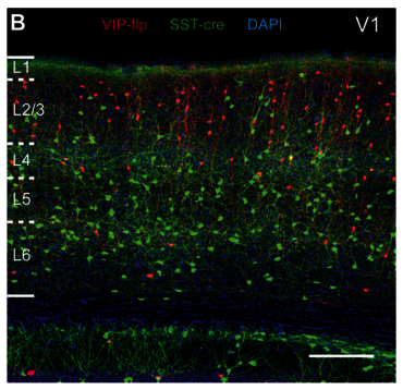
A long-standing question in cortical neuroscience is about the specificity of synaptic wiring motifs between individual identified cell types and how much they differ across different cortical areas. In this study, largely performed by Jenifer Rachel, we compared a translaminar circuit motif between primary somatosensory (S1) and visual (V1) cortices by using paired patch-clamp recordings. We chose these areas since it was previously shown that L4 SST cells in S1 resemble non-Martinotti cells whereas those in V1 are Martinotti cells. With this approach we could make two striking novel observations: (i) L2/3 VIP cells connect with very high probability to both, S1 non-Martinotti cells and V1 Martinotti cells, suggesting that the level of specificity of this connection is rooted in the SST subpopulation as such and not in the different cell types it is composed of; (ii) different forms of short-term plasticity are observed in S1 compared to V1, suggesting that there are sensory modality-specific needs in information processing. Please check out the publication if you are interested to learn more, please check out: https://www.jneurosci.org/content/45/13/e0949242025, or simply for enjoying the beautiful images that the new reporter mouse line provided by our collaborators at the Allen Institute was enabling us to obtain.